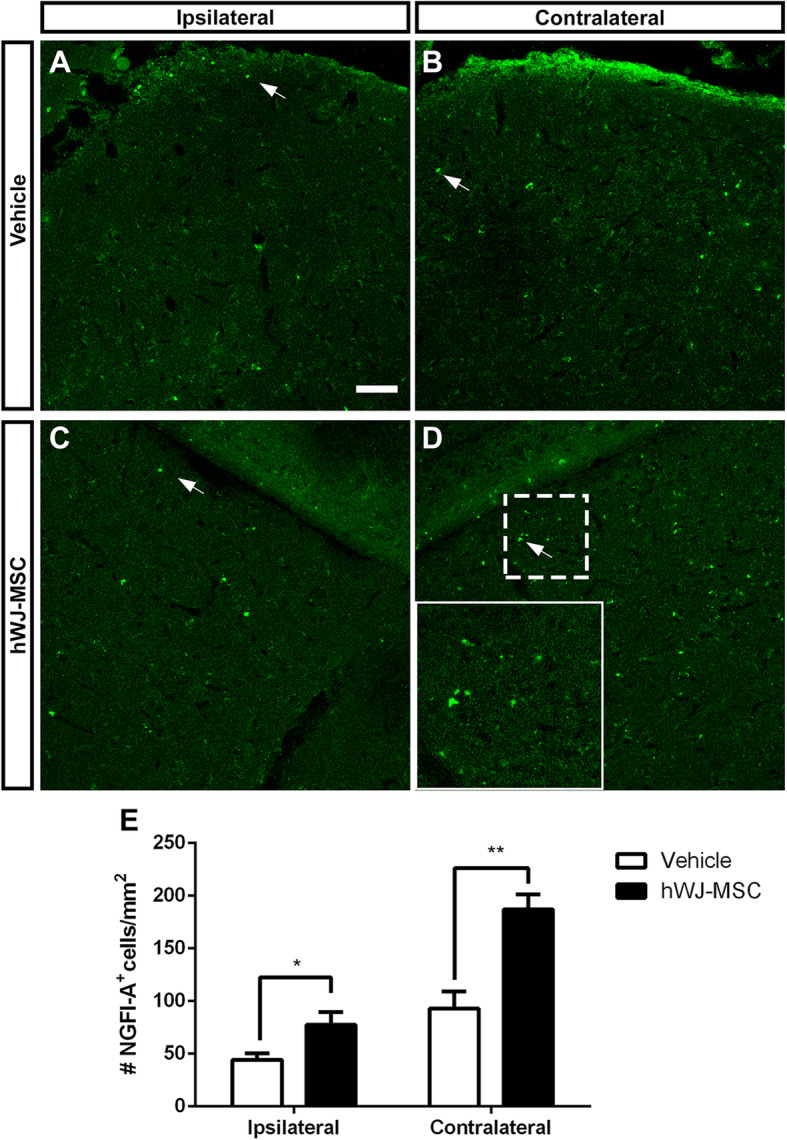
Fig. 6.

RGC terminals activate SC neurons. Images of × 20 z-stack confocal images. NGFI-A immunolabeling to identify activation of SC cells after light exposure in coronal section of ipsi- and contralateral SC 120 days after optic nerve crush and treatment with vehicle (a, b) or hWJ-MSCs (c, d). Inset in d shows higher magnification of the dashed square. e Quantification of the number of cells expressing NGFI-A per mm2 in each group ± SEM. Unpaired parametric t test, *P < 0.05, **P < 0.01. Scale bars 50 μm for images a–d; 23 μm for inset
