Fig. 3.
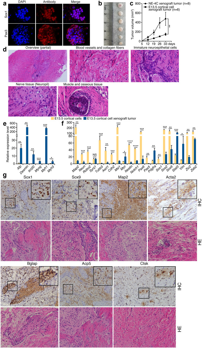
Tumor formation and tissue differentiation by cortical NPCs from E13.5 mouse embryos. a IF staining of neural stemness marker expression in neurospheres formed by cortical NPCs cultured in serum-free medium. Cell nuclei were counter-stained with DAPI. b Xenograft tumors formed by NPCs in nude mice. c Comparison of tumor growth by NE-4C and cortical cells within a same period. Significance of difference in tumor volume between two groups was calculated using two-way ANOVA-Bonferroni/Dunn test. Data are shown as mean ± SEM. *p < 0.05, **p < 0.01, ***p < 0.001, ****p < 0.0001. NS: not significant. d An overview and specific types of tissue differentiation in an HE-stained tumor section. Original objective magnifications: 4× for overview, 40× for specific tissues. e, f Differential expression of genes representing mesendodermal tissue differentiation (e), neural stemness and neuronal differentiation (f) in NPCs and xenograft tumors, as detected with RT-qPCR. Significance of expression change was calculated based on experiments in triplicate using two-tailed Student’s t-test. Data are shown as mean ± SEM. *p < 0.05, **p < 0.01, ***p < 0.001, ****p < 0.0001. NS: not significant. g IHC detection of cell/tissue markers in tumor sections. Below the marker panels are corresponding sections stained with HE. Objective magnification: 20×; insets: 40×

