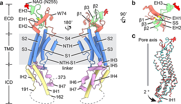
Figure 3: A single hsPANX1 subunit.
a, The disordered IH1-IH2 linker (residues 163–190) and the CTT (residues after 373) are indicated by dashed lines. b, The ECD of single subunit viewed from the extracellular side. SS stands for disulfide bond. c, Superimposition of the subunits of wt-hsPANX1 (red) and N255A-hsPANX1 (cyan) aligned by ECD.

