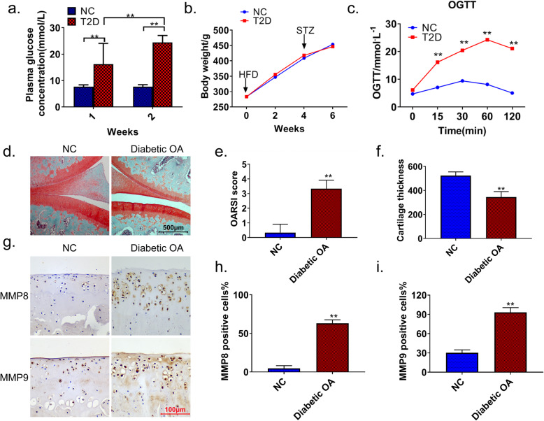
Fig. 1.
Upregulated expression of MMP8 and MMP9 in the knee joint cartilage of diabetic OA rats. a After 4 weeks of a high-fat diet, blood glucose values of the model (STZ) and control (NC) rats were monitored at 1 and 2 weeks after modeling. b The body weights of rats in each group were recorded after 1–6 weeks of OA induction. c OGTT results of STZ and NC rats. d Safranin O-Fast green staining was used to detect articular cartilage lesions in diabetic OA and NC rats. OARSI scores (e) and articular cartilage thickness (f) in diabetic OA and NC rats. g–i IHC showing the expression levels of MMP8 and MMP9 in the joint tissues of diabetic OA and NC rats. Image-Pro Plus was used to quantify the expression. Diabetic OA diabetic osteoarthritis, T2D high-fat diet combined with STZ, NC normal control, OGTT oral glucose tolerance test, MMP matrix metalloproteinase. **p < 0.01

