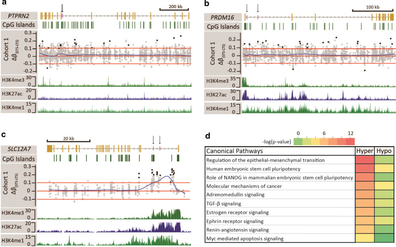
Fig. 2.
Representative STS/LTS differentially methylated regions and ontology analysis. Browser views of the a PTPRN2, b PRDM16, and c SLC12A7 genes. Gene structure is displayed in the first track. Yellow bars represent exons and the grey line introns. Vertical black arrows pointing to short purple bars denote the location of loci that underwent pyrosequencing. Direction of transcription is indicated by arrows on the grey line. The second track displays CpG islands (green bars). Track 3 represent the Δβ(STS—LTS) of all CpGs covered by RRBS 5000 bp upstream and downstream of the gene using data from cohort 1. Each grey circle represents a CpG, and the black circles represent the DMCpG meeting p < 0.01 and |Δβ(STS–LTS)|> 0.1 cutoff. Red lines represent the cutoff line at Δβ(STS–LTS) ± 0.1. The blue line is a smoothed distribution of the data. Tracks 4–6 display peaks of the histone marks H3K4me3, H3K27ac, and H3K4me1, respectively derived from normal kidney. d Heatmap of ontology enrichments for a top group of pathways derived from genes linked to the 5929 DMCpGs between STS and LTS ccRCCs

