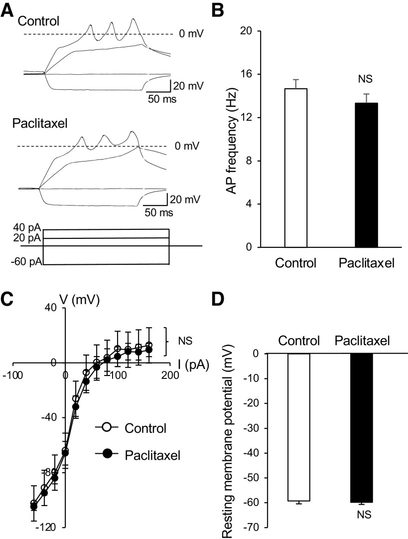
Fig. 2.
Lack of effects by paclitaxel on membrane electrophysiological properties of Merkel cells. A: sample traces show membrane responses and action potentials recorded from a Merkel cell. Shown are recordings before (control; top) and following the bath application of 1 µM paclitaxel for 30 min (bottom). Membrane responses were elicited by injections of current steps each at 20 pA. B: summary data of action potential (AP) frequency in Merkel cells of control (n = 6) and following the paclitaxel application (n = 6). C: voltage-current relationship of membrane responses in Merkel cells of the control (n = 6) and following the bath application of paclitaxel for 30 min (n = 6). D: summary data of resting membrane potentials of Merkel cells in the control (open bar, n = 6) and following the bath application of paclitaxel for 30 min (n = 6). Data represent means ± SE. NS, not significantly different, two-way ANOVA with Bonferroni post hoc tests or paired Student’s t test.

