Fig. 5.
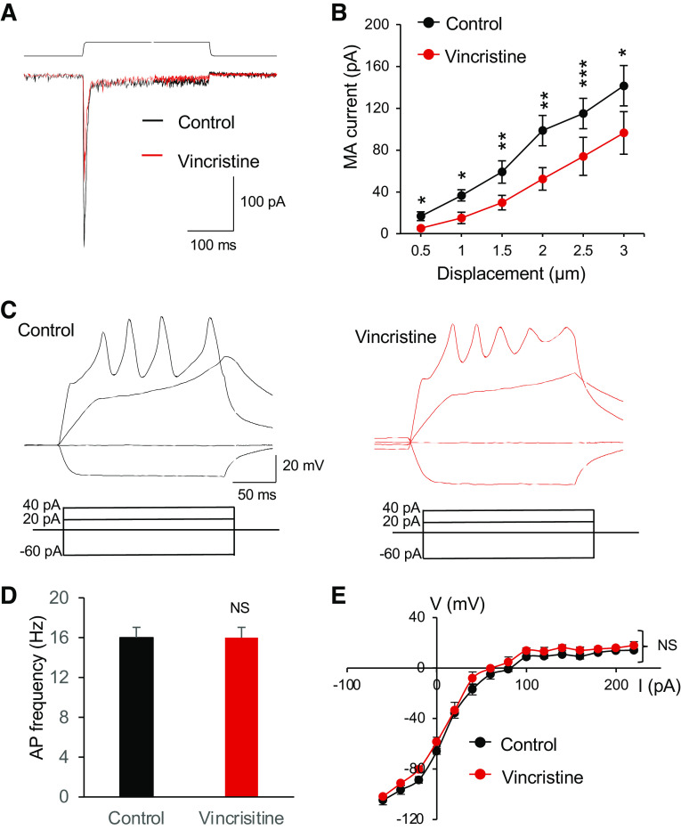
Effects of the microtubule destabilizer vincristine on mechanically activated currents and membrane electrophysiological properties of Merkel cells. A: overlay sample traces show mechanically activated (MA) currents from a Merkel cell before (control, black) and following the bath application of 100 µM vincristine for 30 min. The mechanical stimulation was a 3-µm membrane displacement indicated on the top of MA currents. B: summary data of MA currents recorded from Merkel cells before (control, n = 6) and following the bath application of 100 µM vincristine for 30 min (n = 6). The MA currents were evoked by membrane displacements at 0.5, 1, 1.5, 2, 2.5, and 3 µm. C: sample traces show membrane responses and action potentials recorded from a Merkel cell in the control (left) and following the bath application of 100 µM vincristine for 30 min (right). Membrane responses were elicited by injections of current steps each at 20 pA. D: summary data of action potential (AP) frequencies in Merkel cells in the control (n = 6) and following the bath application of 100 µM vincristine for 30 min (n = 6). E: voltage-current relationship of membrane responses in Merkel cells of the control (n = 6) and following the bath application of 100 µM vincristine for 30 min (n = 6). Data represent means ± SE. NS, not significantly different; *P < 0.05, **P < 0.01, ***P < 0.001, two-way ANOVA with Bonferroni post hoc tests or paired Student’s t test.

