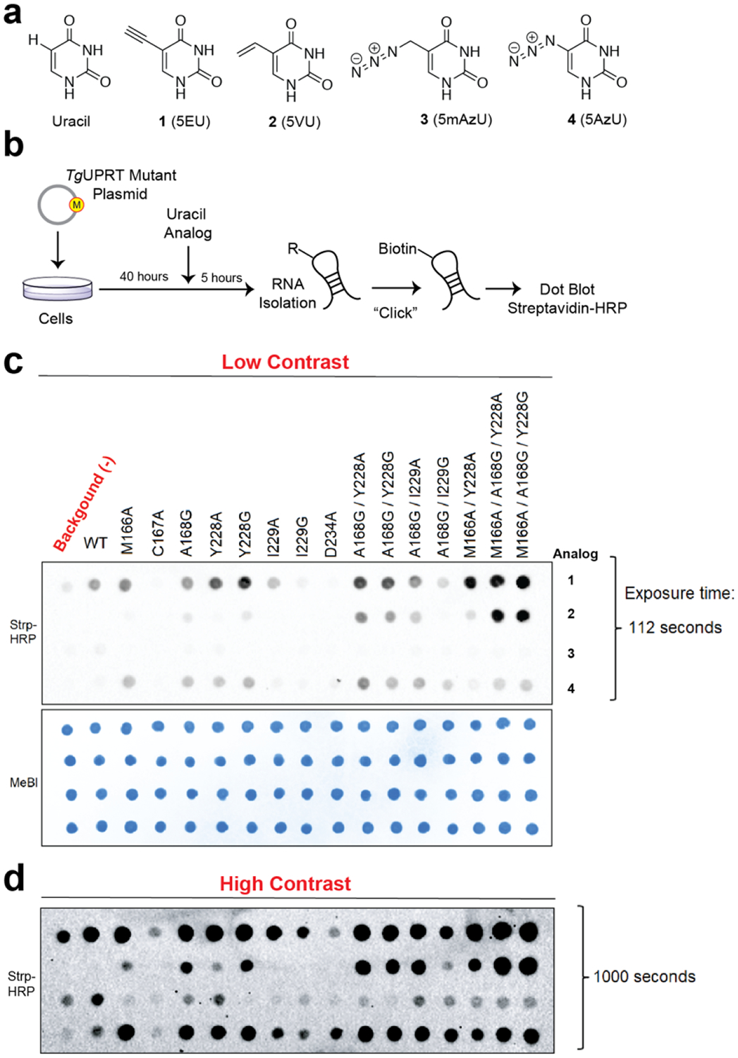
Figure 2. In-cell screening of TgUPRT mutants matched with bioorthogonal analogs.

a. Chemical structures of uracil and uracil analogs used herein. b. Schematic of in-cell screening experiments. HEK293T cells were transfected with TgUPRT plasmids containing various mutations. Uracil analogs were added at 200 μM and incubated for 5 hours. Following RNA isolation, biotinylation was performed and incorporation of analog was determined by streptavidin dot blot. c. Dot blot screening for RNA incorporation of four different uracil analogs by fifteen TgUPRT mutants. d. Longer exposure of the dot blot shown in panel c. MeBl = methylene blue staining served as loading control.
