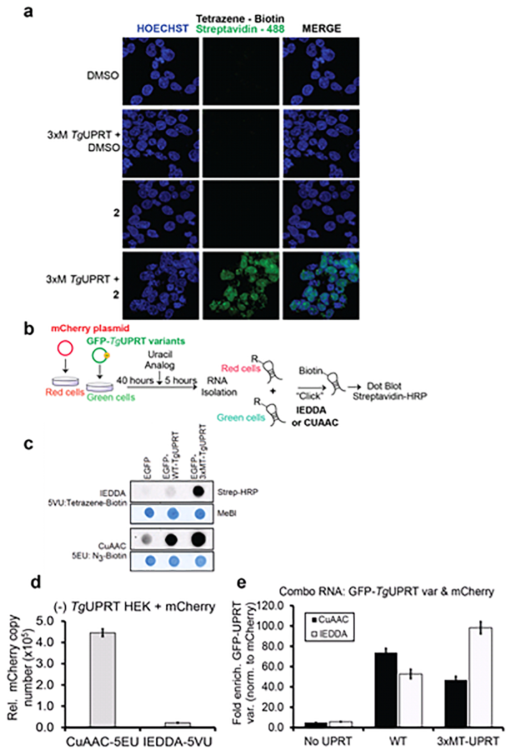
Figure 4. Characterizing the stringency of the 2 (5VU)-mutant TgUPRT pair.

a. Microscopy analysis RNA incorporation of 2 in a mutant TgUPRT-dependent manner. Cells were transfected with TgUPRT variants and incubated with 2 at 1 mM final concentration for 24h. 2 incorporation was imaged using two-step labeling: IEDDA using Tetrazine-biotin then followed by Alexa488-streptavidin. b. Schematic of experiment to assess specificity of 2 in cells transfected with mCherry (No UPRT) or GFP, GFP-TgUPRT variants treated with both 200 μM 1 and 400 μM 2. c. Dot blot analysis of RNA isolated from 1-2 treated cells underwent either CuAAC or IEDDA click reaction. Streptavidin-HRP was used for assessment of biotin levels resulting from clicked RNA, and methylene blue (MeBl) staining served as loading control. d. QPCR analysis of enriched CuAAC- or IEDDA-biotinylated mCherry cDNA from (−)TgUPRT HEK293T cells. e. QPCR analysis of enriched CuAAC or IEDDA-biotinylated GFP cDNA from (−)TgUPRT (No UPRT), WT-, and 3xMT-TgUPRT (M166A/A168G/Y228A) transfected HEK293T cells
