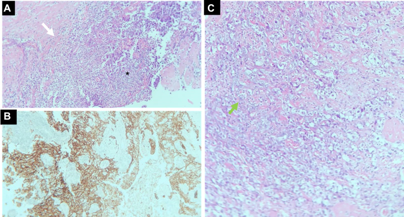
Figure 5.
(A–C) Tumor histology with the diagnosis of medullary carcinoma. Microscopy at lower magnification demonstrates undifferentiated cells with syncytial growth, intense inflammatory infiltrate with a predominance of T lymphocytes (black asterisk), and invasion of the capsule (white arrow) (A). The immune profile (pancytokeratins) shows staining the epithelial cells with the tumor markers AE1 and AE3 (B). In a greater augmentation, foamy histiocytes are observed between the inflammatory process (green arrow) (C).

