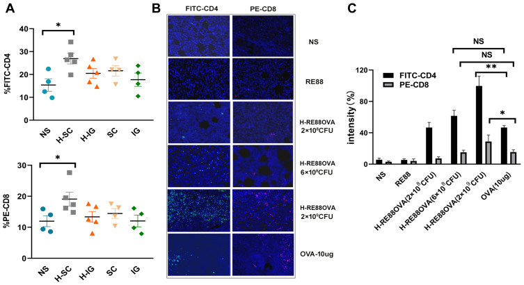
Figure 4.
Infiltration of EG7-OVA tumor tissue by lymphocytes. (A) Percentages of intratumoral CD4+/CD8+ T lymphocytes. Lymphocyte infiltration of tumor tissue in mice immunized subcutaneously or intragastrically with 2 × 10⁸ CFU of non-inactivated or H2O2-inactivated RE88-pVLT33-OVA. Data are pooled from two independent experimental replicates. (B) Lymphocyte infiltration of tumor tissue in mice immunized subcutaneously with various types of vaccine. CD4-positive T cells were labeled with FITC (green), CD8-positive T cells were labeled with PE (red), and nuclei were labeled with DAPI (blue). (C) Mean fluorescence intensity indicating the extent of tumor infiltration with CD4-positive and CD8-positive T cells. Data are presented as mean ± SEM (n ≥ 3). *P < 0.05, **P < 0.01, NS, P > 0.05. H-IG: Intragastric administration of H2O2-inactivated RE88-pVLT33-OVA; H-SC: subcutaneous administration of H2O2-inactivated RE88-pVLT33-OVA; IG: intragastric administration of non-inactivated RE88-pVLT33-OVA; SC: subcutaneous administration of non-inactivated RE88-pVLT33-OVA. NS: Normal saline (negative control); RE88: unmodified strain; H-RE88OVA: subcutaneous administration of H2O2-inactivated RE88-pVLT33-OVA (3 doses); OVA-10 µg (positive control).

