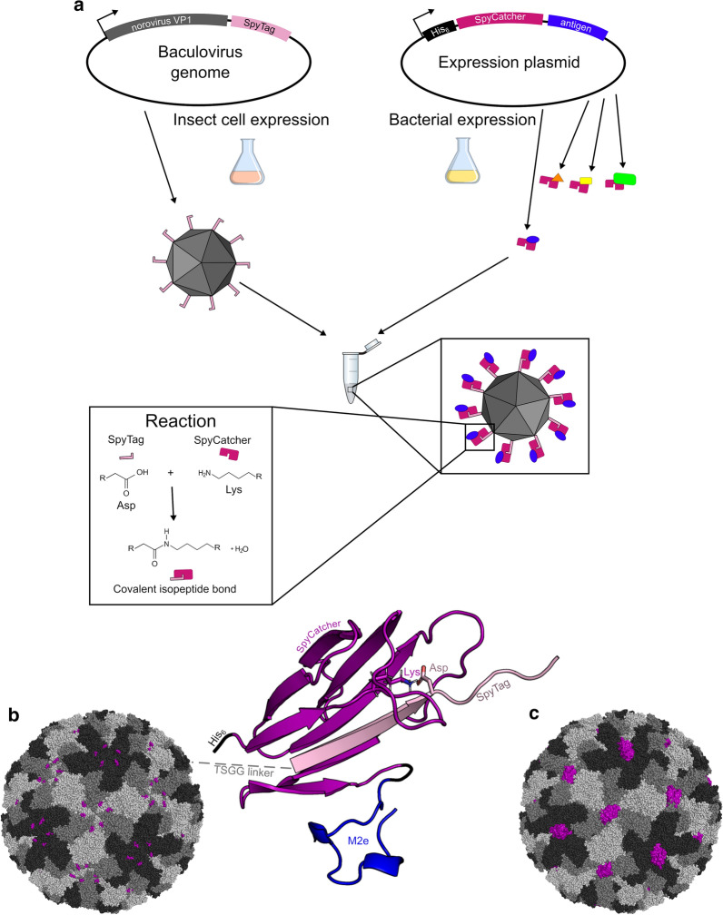
Fig. 1.
Overview of SpyTag-norovirus-like particle vaccine design. a When SpyTagged norovirus VP1 protein is expressed in insect cells, the proteins spontaneously assemble to form noro-VLPs with SpyTag peptides protruding from their surface. Then, this SpyTag-norovirus-like particle is mixed with a separately produced SpyCatcher-antigen fusion protein (depicted here as blue/orange/yellow/green antigen on purple SpyCatcher), and a biologically irreversible covalent bond forms in between. b The noro-VLP is formed by 180 VP1 proteins, whose C-termini (purple) protrude from the surface in clusters of five or six. SpyTag (light purple) was genetically fused to the C-terminus of noro-VP1 via a 4-residue linker. On a different scale, the panel shows a conjugated SpyCatcher (dark purple), fused to M2e (blue) by its C-terminal. c One SpyCatcher bound per cluster of SpyTags is shown for size comparison. The fusion protein models in b and c were constructed with the SynLinker web application [46] (maintenance ceased since) and visualized with PyMOL [47]. The models are based on the following RCSB PDB structures: norovirus: 1IHM, SpyCatcher: 4MLS, M2e: 4N8C

