
Figure 2. a–f.
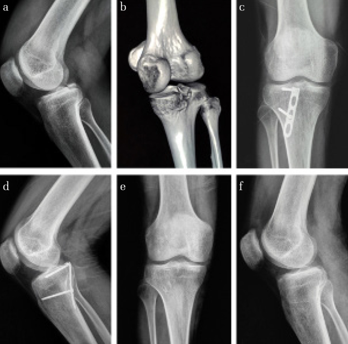
(a) Lateral radiograph of the knee (nonstress view) showing the avulsed fragment from the tibia. (b) Three-dimensional computed tomography scan showing the avulsed fragment of the tibia. (c and d) Radiographs showing bone union 3 months after surgery. (e and f) Photographs of the 3.5-mm LCHP plate removed 1 year after surgery