
Fig. 1.
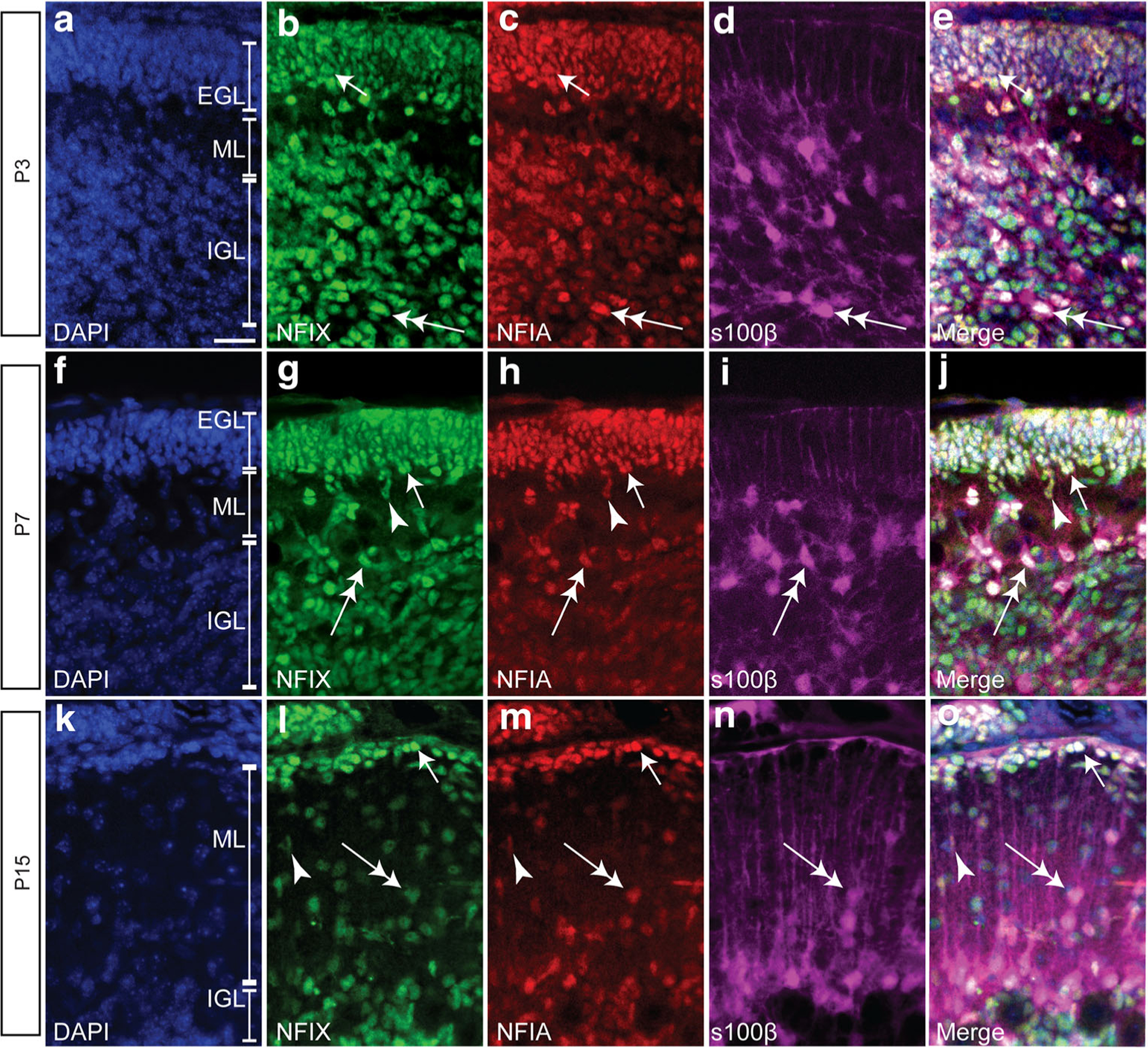
NFIX and NFIA are co-expressed by various cellular populations in the developing mouse postnatal cerebellum. Sagittal cerebellar sections showing the nuclear marker DAPI (blue), NFIX (green), NFIA (red) and s100β expression (magenta) in P3 (a–e), P7 (f–j) and P15 (k–o) wild-type mice. NFIX and NFIA are co-expressed by GNPs within the external granule layer (EGL; arrows in b–e, g–j, l–o). NFIX and NFIA are also co-expressed by s100β-positive glial cells (double-headed arrows in b–e, g–j, l–o). These transcription factors are also expressed by cells leaving the EGL (arrowheads in g–j, l–o); these are likely immature neurons migrating to the internal granule layer (IGL). ML molecular layer. Scale bar (in A) = 20 μm