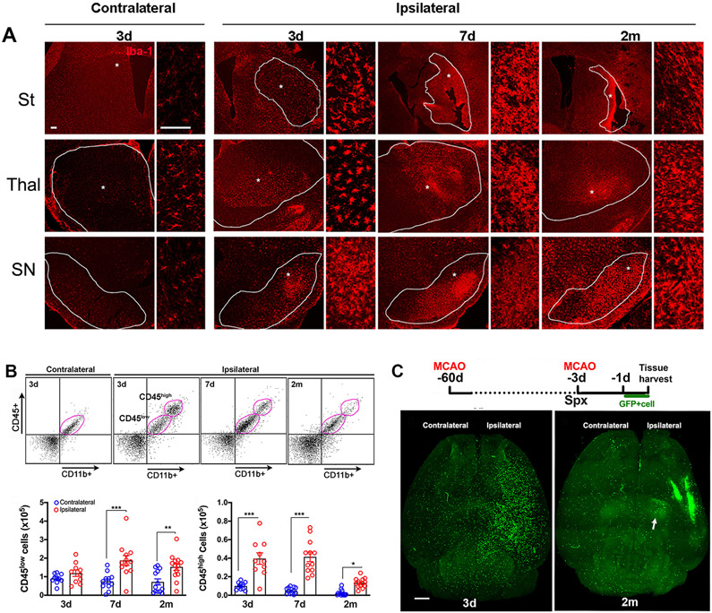
Figure 6.
Mononuclear phagocyte localization and trafficking. A, Iba-1 immunofluorescence. Dotted area in striatum (St) indicates infarct zone. Dotted lines in the thalamus (thal) and substantia nigra (SN) demarcate the sub-regions. * high magnification. Scale bar, 50μm. B, Representative flow cytometry plots and qunatification for CD45low and CD45high subsets in the pose-stroke brain. n=10-13/timepoint. Values are expressed as mean ± s.e.m. Two way ANOVA, Fisher’s LSD test, *, **, *** p<0.05, 0.01, 0.001 versus contralateral. C, Experimental timeline. GFP+ splenocytes was adoptively transferred to asplenic (Spx) mice one day prior to sacrifice. Representative horizontal view images from cleared whole brain at 3d and 2 m post-stroke. Scale bar,1mm.

