
Figure 8. SCIII2+IV interface in V. radiata.
(A) General orientation of SC III2+IV in ribbon representation viewed from the membrane. Approximate position of the inner mitochondrial membrane is shown. Sites 1 (S1) and 2 (site 2) of the supercomplex interface are marked in dashed circles. (B) Detailed view of the protein-protein interaction in site 1 (Pro31 of VrQCR8 and Trp59 of VrCOX2) with the interacting atoms shown in stick representation. Note that interacting residues of site two appear in stick in the background. (C) Detailed view of the protein-protein interaction in site 2 (Pro19-Lys20 of QCR6, Gln70-Phe72 of COX4, Leu26 of QCR6, Leu51 of COX5c) with the interacting atoms shown in stick representation.
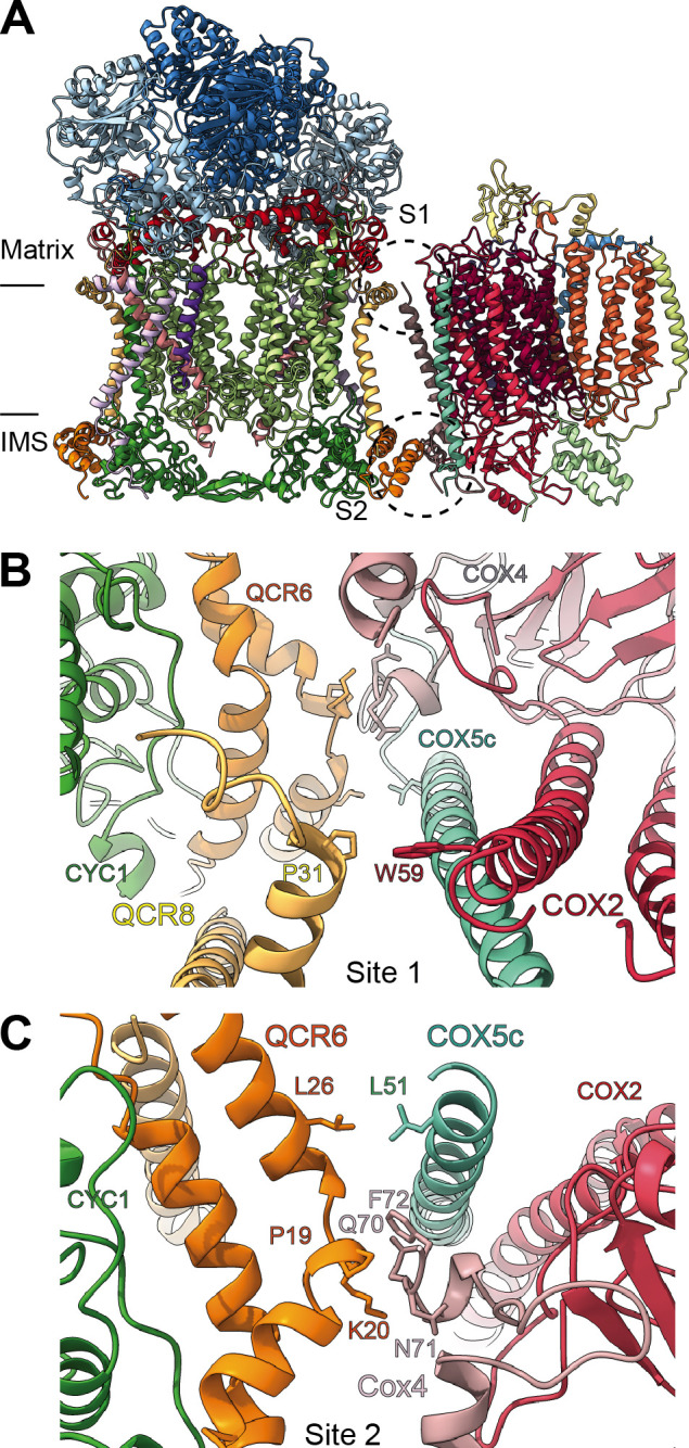
Figure 8—figure supplement 1. Differences in SC III2+IV interactions between V. radiata and S. cerevisiae (PDB: 6HU9).

The supercomplexes are aligned by V. radiata’s COB and CYC1 and shown in surface representation. (A-B) V. radiata (A) and S. cerevisiae (B) SC III2+IV viewed from the membrane. (C-D) Superposed V. radiata (green) and S. cerevisiae (pink) viewed from the matrix (C) or the intermembrane space, IMS (D). (E) Cyt c (yellow) docked onto CIII2 and CIV in superposed V. radiata (green) or S. cerevisiae (pink) SC III2+IV viewed from the IMS. Distance between the CIII2- and CIV-bound cyt c in the V. radiata (Vr) and S. cerevisiae (Sc) supercomplexes shown. Cyt c was docked based on the S. cerevisiae CIII2-bound cyt c (1KYO) and the bovine CIV-bound cyt c (5IY5). CIII2-bound cyt c was docked onto V. radiata CIII2 by aligning CYC1. CIV-bound cyt c was docked onto V. radiata and S. cerevisiae CIV by aligning COX2. Distance was measured edge-to-edge of the heme conjugated ring systems.