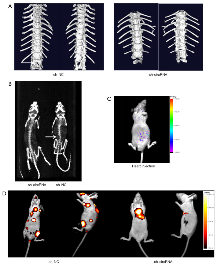
Figure 7.
Knockdown of hsa_circ_0006571 suppressed SM in vivo. (A,B) Representative micro-computed tomography and X-ray scans showing local osteolytic lesions (arrow) in the affected vertebrae of tumorigenic nude mice. (C) After left ventricular injection, the tumor cells were distributed all over the body, according to images of luciferase signals. (D) Images of luciferase signals in nude mice inoculated in the left ventricle with luciferase-labeled tumor cells. SM, spinal metastasis.

