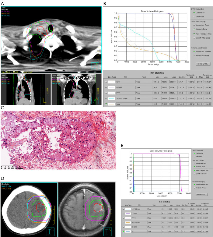
Figure 2.
Thoracic and intracranial radiotherapy plans. (A) Contouring of lesions and isodose curves in thoracic radiotherapy. (B) Dose-volume histogram (DVH) of the thoracic radiotherapy plan. (C) Hematoxylin and eosin (HE) staining of a surgically removed intracranial lesion, original magnification ×100. (D) Postoperative radiotherapy plan for brain metastasis. (E) DVH of the intracranial radiotherapy plan.

