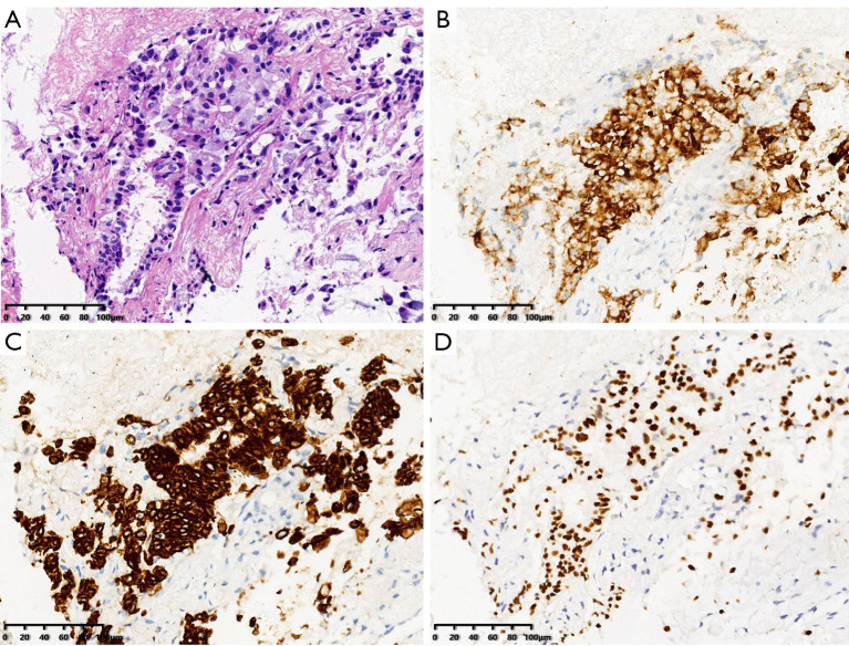
Figure 3.
Pathologic findings of a thoracic metastatic biopsy lesion. (A) HE staining, original magnification ×200. (B) ALK rearrangement detected by immunohistochemistry (IHC), original magnification ×200. (C) Cell keratin 7 (CK7) expression detected by IHC, original magnification ×200. (D) Expression of thyroid transcription factor (TTF)-1 by IHC, original magnification ×200.

