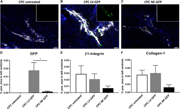
FIGURE 7.
GFP expression and CPC engraftment 1 week after transplanting CPCs to the left ventricle near the left anterior descending artery. (A–C) Typical examples of expression of GFP (green), β1-integrin (red), and Collagen-1 (white) in mouse hearts injected with (A) unmodified CPCs, (B) LV-GFP transduced CPCs or (C) GFP nucleofected (NF) CPCs. Inset: GFP expression alone. Scale bars represent 100 μM. (D–F) Expression of GFP, β1-integrin, and Collagen-1 in mice injected with unmodified CPCs (n = 4), lentivirally transduced CPCs (n = 4) or nucleofected CPCs (n = 5), measured as% positive area of total left ventricular area. * indicates P < 0.05.

