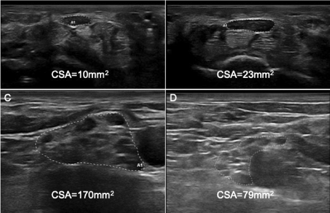
Fig. 2.
Nerve ultrasound of median nerve at wrist and brachial plexus in supraclavicular space. Panel a shows a severe CTS (grade 4, accordingly to Padua’ scale) in an ATTRv woman, CSA was within normal limit (10mm2), while in b an idiopathic CTS of the same severity showed a markedly enlarged CSA (23 mm2). In C and D right brachial plexus at supraclavicular space are showed. In c is reported the enlarged plexus of an ATTRv patient (with late-onset Val30Met mutation) with polyneuropathy, in d the normal plexus of the pre-symptomatic son of the same patients. CSA Cross sectional area

