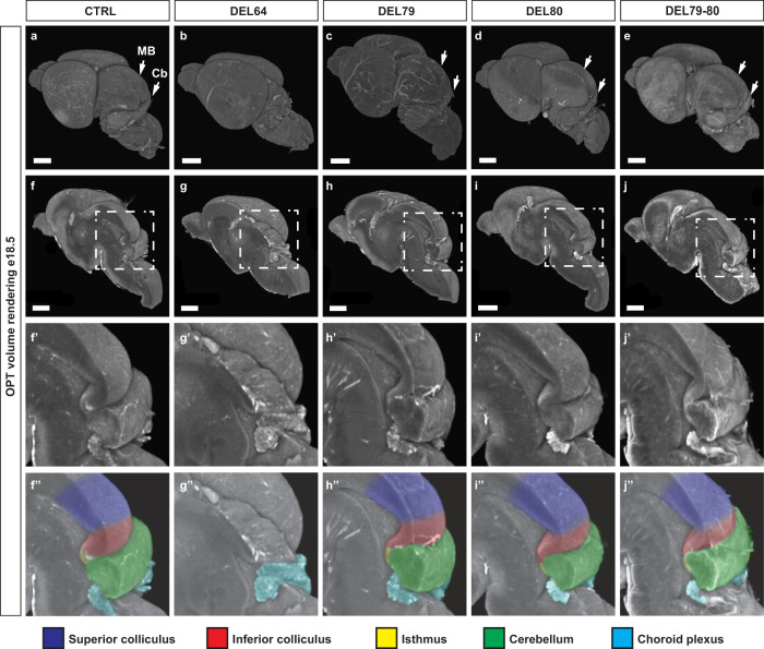
Fig. 2. One main enhancer is required for Fgf8 expression in the MHB.
a–j” OPT generated volume renderings of e18.5 brains from control (a, f, f’, f”), DEL64 (b, g, g’, g”), DEL79 (c, h, h’, h”), DEL80 (d, i, i’, i”) and DEL79-80 (e, j, j’, j”) mutants. Signal is based on tissue autoflourescence. Control brains, DEL79, DEL80 and DEL79-80 mutants display a well-developed midbrain and cerebellar anlage while DEL64 brains display severe hypoplasia of midbrain and cerebellum. f–j Midsagittal digital dissection reveal complete loss of all MHB derived structures in the DEL64 mutant. f’–j” Close-up of boxed area in (f–j). Brains have been pseudocolored in (f”–j”): dark blue, superior colliculus; red, inferior colliculus; yellow, isthmus; green, cerebellum; light blue, choroid plexus. Scale bar in (a–j) is 1 mm. MB, midbrain; Cb, cerebellum.

