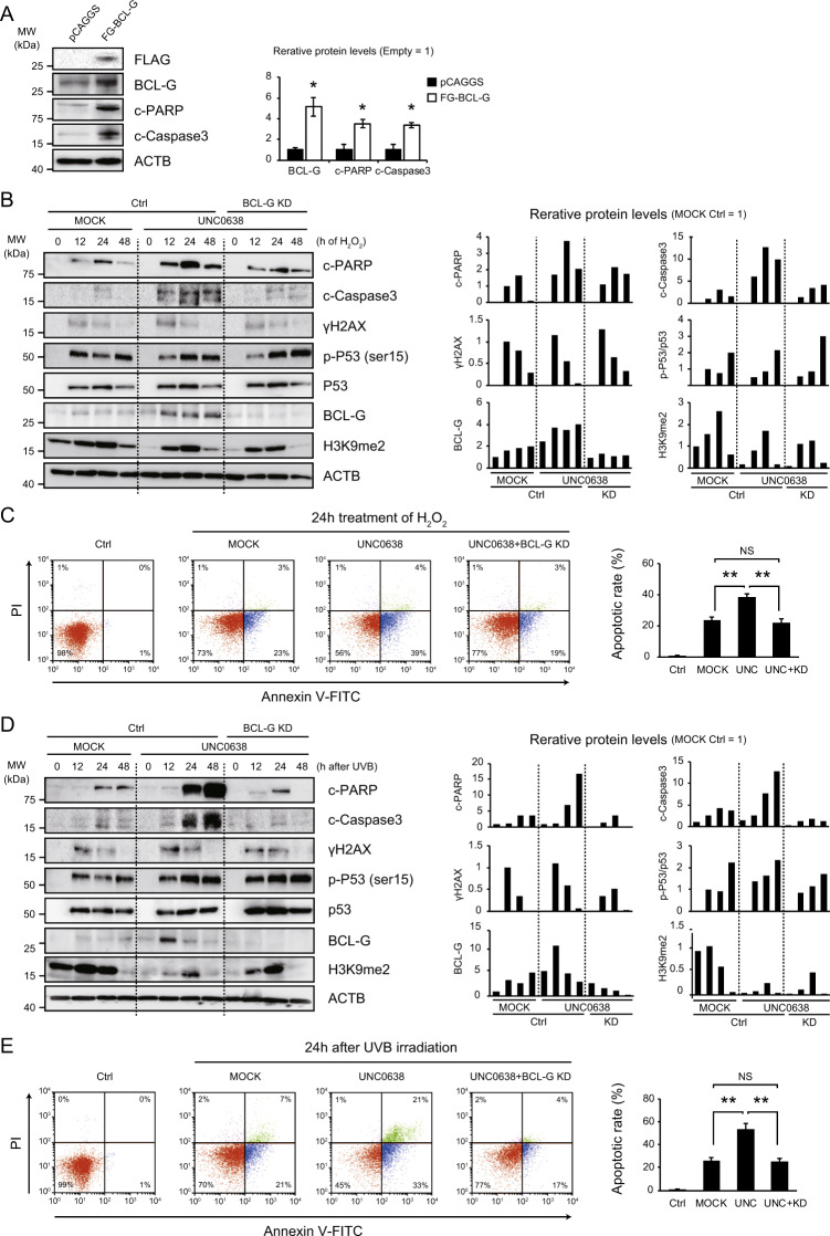
Fig. 4. BCL-G is involved in apoptosis during the DNA damage response in hepatocytes.
A BCL-G overexpression induces apoptotic changes in normal human hepatocytes. Representative immunoblot images and relative expression levels of the proteins are shown (each, n = 3; *p < 0.05, Student’s t test). B, D The G9a inhibitor UNC0638 (5 µM) promotes apoptosis in hepatocytes after treatment with hydrogen peroxide (H2O2, 2 mM) or UVB irradiation (300 J/m2) with an increase in BCL-G expression. Bcl-G knockdown (KD) suppresses the excessive induction of apoptosis caused by a G9a inhibitor. Right graphs show quantification of relative protein levels. C, E Representative dot-plot diagrams showing the percentage of the hepatocyte lines stained with Annexin V-FITC and propidium iodide (PI) after 24 h of treatment with H2O2 or UVB irradiation. The results of three independent experiments are shown in the right graph (**p < 0.01, NS not significant, Student’s t test).

