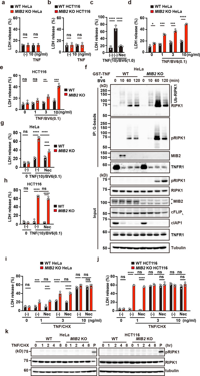
Fig. 6. RIPK1 kinase activity-dependent and -independent apoptosis are enhanced in MIB2 KO cells.
a, b, d, e WT and MIB2 KO cells were unstimulated, stimulated with TNF (10 ng/ml) (a, b), or the indicated concentrations of TNF and low concentrations of BV6 (0.1 μM) (d, e) for 8 h. Cell death was determined by the LDH release assay. Results are mean ± SD of triplicate samples. c WT HeLa cells were stimulated with TNF (10 ng/ml)/high concentrations of BV6 (1.0 μM) in the absence or presence of Nec-1s (20 μM) for 8 h. Results are expressed as in a. f WT and MIB2 KO HeLa cells were stimulated with GST-TNF (1 μg/ml)/BV6 (0.1 μM) for the indicated times, and the TNFR-containing complex was precipitated and analysed as in Fig. 5a. g–j WT and MIB2 KO cells were unstimulated or stimulated with the indicated concentrations of TNF and low concentrations of BV6 (0.1 μM) (g, h) or CHX (2.5 μg/ml) (i, j) in the absence or presence of Nec-1s (20 μM). Results are expressed as in a. Unpaired two-tailed Student t-test (a, b, d, e), one-way ANOVA with Tukey’s multiple comparison test (c), or two-way ANOVA with Bonferroni’s multiple comparison test (g–j). *P < 0.05; **P < 0.01; ***P < 0.001; ****P < 0.0001; ns, not significant. k WT and MIB2 KO cells were stimulated with TNF (1 ng/ml)/CHX (2.5 μg/ml) for the indicated times. Cell lysates were analysed by immunoblotting with the indicated antibodies. P indicates positive control lysates of MIB2 KO HeLa cells stimulated with TNF/BV6 (0.1 μM) for 2 h. All results are representative of at least two independent experiments.

