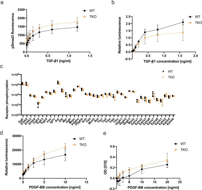
Fig. 5. TGF-β and RTK signaling appear unaltered in Lrig-null MEFs.
a Wild-type (WT) and Lrig-null (TKO) MEFs were stimulated with various concentrations of TGF-β1 for one hour and then analyzed for nuclear phospho-Smad3 (pSmad3) by an immunocytofluorescence assay. b Wild-type and Lrig-null MEFs were transiently transfected with the TGF-β reporter plasmid p(CAGA)12MLP-Luc followed by treatment of the cells with the indicated concentrations of TGF-β1 for three hours. Cell lysates were then analyzed for luciferase activity. Shown is the relative luminescence on an arbitrary scale. c Wild-type and Lrig-null MEFs, cultivated under standard cell culture conditions in 10% FBS, were lysed and analyzed for RTK phosphorylation levels with a phospho-RTK array kit (R&D Systems). d Wild-type and Lrig-null MEFs stably expressing the MAPK reporter gene ELK1/SRF-luc were treated with different concentrations of PDGF-BB for four hours and then lysed and analyzed for luciferase activity. e Wild-type and Lrig-null MEFs were cultivated in FBS-free medium containing different amounts of PDGF-BB for 48 h. Thereafter, the relative cell numbers were determined using an MTT assay. Shown are the OD values of treated MEFs normalized to those of untreated MEFs. a, b, d and e The plotted values represent the means of four biological replicates, each with three experimental repeats. c The plotted values represent the means of four biological replicates, each with one experimental repeat. Error bars represent the standard deviations of the means from four biological replicates.

