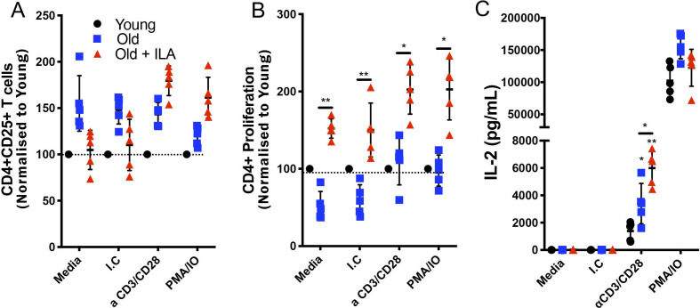
Figure 4.
CD4 +T cells from persons with interstitial lung abnormalities (ILA) display high proliferative capacity compared with old control group. Enriched T cells were cultured 48 hours with polyclonal stimuli using either monoclonal antibodies anti-CD3 and CD28 (both at 1 µg/mL) or phorbol myristate acetate (PMA) 50 nM/ionomycin (IO) 2 ng/µL, culture medium (Media) and isotype control (I.C) to monoclonal antibodies were used as negative controls. Cells were recovered and prepared to flow cytometry and culture supernatants were used to determine interleukin (IL)-2 by ELISA. By flow cytometry, CD3+CD4+cells were delimited and the expression of CD25 (for activation) and Ki-67 (for proliferation) were evaluated (panel A and B, respectively); the level of activation and proliferation displayed for CD4+T cells from young group was considered as 100% (black dot line). Panel C: IL-2 released by stimulated T cells was measured by ELISA in the culture’ supernatants. Graphs show individual value mean±SD (n=5 per group). *p<0.05 **p<0.01. Statistical analysis was performed with one-way analysis of variance test and Dunnett as post hoc analysis (ELISA data), and multiple t-tests with a Holm-Sidak to adjustment for multiple comparisons (flow cytometry data). Asterisks indicate comparison of the three groups. When the asterisk is on a line, indicates, the comparison between old control group and old+ILA. M-CSF, macrophage colony-stimulating factor; TNF, tumor necrosis factor.

