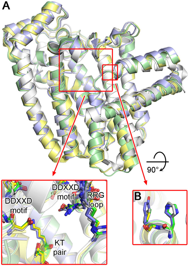
Figure 11.

Structures of trypanosomatid FPPS. (A) Superimposed structures of T. cruzi (green; PDB ID: 4DWG), T. brucei (light purple; PDB ID: 4RXD), L. major (yellow; PDB ID: 4JZX) and human FPPS (white; PDB ID: 4H5E). Conserved residues of the active site are displayed in the inset. (B) Aromatic residues of the allylic substrate site hydrophobic pocket.
