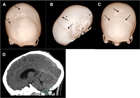
Figure 2.

(A) An 8‐year‐old XLHR patient. Three‐dimensional CT skull bone window reconstruction. Complete closure of the sagittal and the right coronal sutures. The left coronal suture is patent but narrow. The patient also shows a congenital parietal foramina. (B) A 6‐year‐old XLHR patient. Three‐dimensional CT skull bone window reconstruction. Complete closure of the sagittal suture and both lambdoid sutures. (C) A 12‐year‐old XLHR patient. Three‐dimensional CT skull bone window reconstruction. Patent sagittal and coronal sutures. All very narrow. (D) An 8‐year‐old XLHR patient. Head CT sagittal slice parenchyma window showing a 6.8‐mm descent of the cerebellar tonsils. Continuous black arrows indicate sagittal suture. Discontinuous black arrows indicate coronal sutures. Continuous white arrow indicates lambdoid suture. Asterisks indicate closed sutures.
