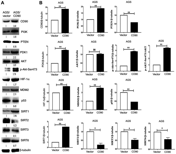
Figure 5.
Western blotting analysis of the PI3K/AKT/HIF-1α signaling pathway. (A) Levels of PI3K, PTEN, PDK1, AKT, p-AKT-Ser473, HIF-1α, MDM2, P53, SIRT2, SIRT3 and SIRT6 were assessed via western blotting. Left, AGS-vector cells; right, AGS-CD90 cells. (B) Densitometric analysis of the western blotting results. *P<0.05, **P<0.01. HIF-1α, hypoxia-inducible factor 1α; SIRT, Sirtuin; Vector, gastric cancer cells transfected with pEGFP-N1; CD90, gastric cancer cells transfected with pEGFP-N1/CD90.

