Figure 6.
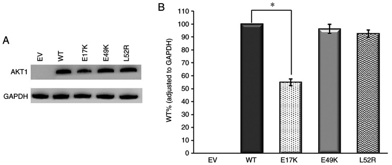
Western blot analysis of AKT1 variants. (A) Immunoreactive AKT1 and GAPDH protein bands and (B) AKT1 WT, E17K, E49K and L52R protein levels. GAPDH was used as the internal control for gene expression. Anti-DDK monoclonal antibody was used for AKT1 detection. n=3. *P<0.02 vs. respective WT. EV, empty vector; WT, wild-type.

