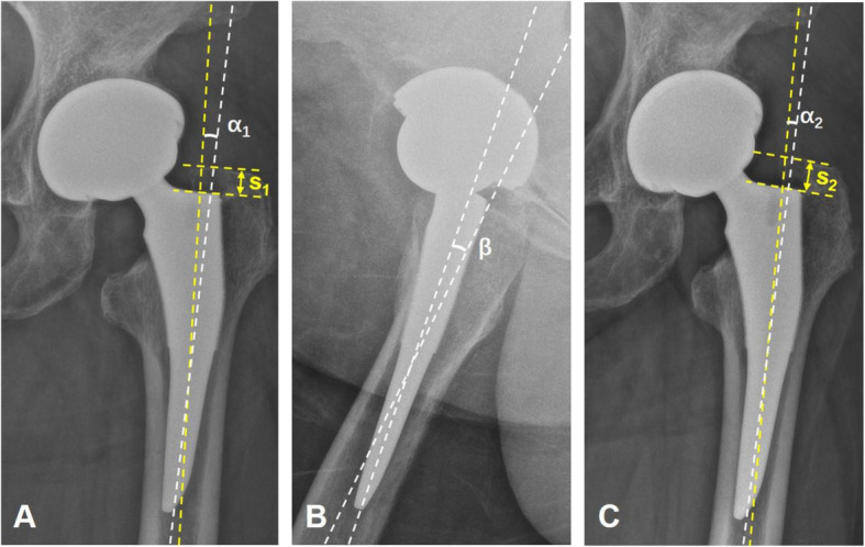
Fig. 2.

a–c A representative image for type I fit. This patient was a 62-year-old woman who underwent THA at age 60 using a short tapered stem, which is a type I fit. a A postoperative AP radiograph of her left hip. b A postoperative lateral radiograph of the hip. C A 6-month postoperative AP radiograph. The following parameters were measured: (α1) CSA, which represents the angle between the stem axis and the femur axis at the first follow-up; (α2) CSA, which represents the angle between the stem axis and the femur axis at the last follow-up; (s1) distance between the major trochanter apex and the stem shoulder perpendicular to the femoral stem axis at the first follow-up; (s2) distance between the major trochanter apex and the stem shoulder perpendicular to the femoral stem axis at the last follow-up; and (β) SSA, which represents the angle between the stem axis and femur axis at the first follow-up. In the AP radiograph, if α1/α2 > 0, it illustrates the stem alignment is valgus, and if α1/α2 < 0, it illustrates the stem alignment is varus. In the lateral radiograph, if β > 0, it illustrates the stem alignment is retroverted, and if β < 0, it illustrates the stem alignment is anteverted
