
Figure 4.
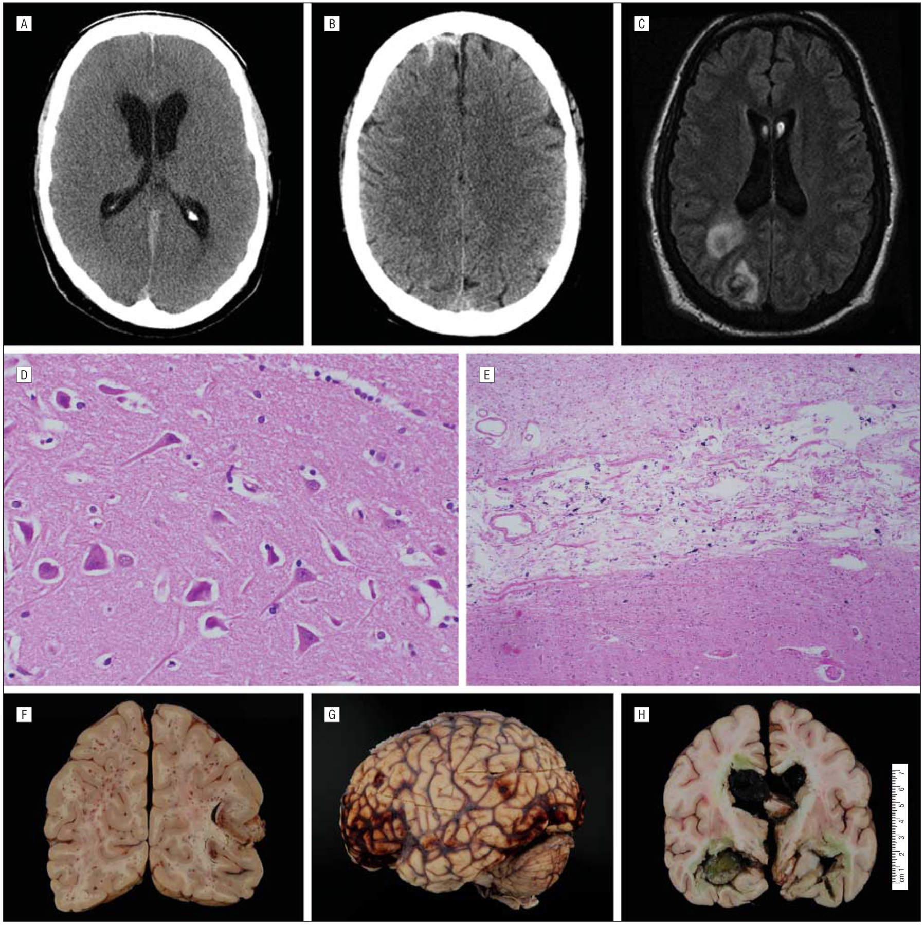
Diagnostic results of adult patients who received extracorporeal membrane oxygenation. The figure shows parafalcine subarachnoid hemorrhage and hydrocephalus on axial-view head computed tomography (A), diffuse subarachnoid hemorrhage on T1-weighted magnetic resonance imaging (B), and septic cerebral emboli on axial-view magnetic resonance imaging (C), which enhances with gadolinium-contrast; acute ischemic cell changes (“red dead neurons”) (D) and microscopic subacute ischemic thalamic infarction on histopathological sectioning (E); and diffuse petechial hemorrhages (F), subarachnoid hemorrhage (G), and massive intraventricular hemorrhage on gross pathological examination (H).