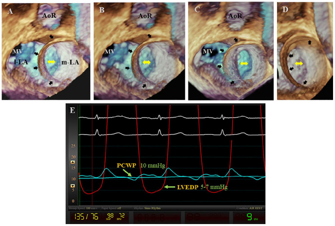
Figure 2.
(A-D) These 3-dimensional transesophageal echocardiogram (TEE) views from the left atrium (LA) at different orientations demonstrate a well-defined curvilinear membrane (wide arrows) extending from the inferomedial to the superior regions of the atria and therefore dividing the LA into a funnel and C-shaped medial LA (m-LA) cavity with a large distal and oval shape opening (double-headed arrows) and an even larger and also C-shaped lateral LA (l-LA) cavity. Note that the membrane appears to attach to the anteromedial and posteromedial, but not to the inferomedial aspects of the atrial wall. (E) This simultaneous recording of the pulmonary capillary wedge pressure (PCWP) and left ventricular pressure demonstrate a mean PCWP of 10 mm Hg and a left ventricular end-diastolic pressure (LVEDP) of 5 to 7 mm Hg and, therefore, a differential gradient of 3 to 5 mm Hg. MV, mitral valve; AoR, aortic root.

