
Figure 6. SG SUMOylation Is Dysregulated by Poly(PR)50 and Ameliorates ALS Phenotype in Flies.
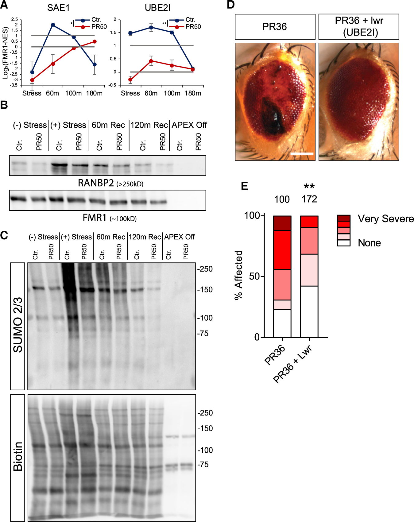
(A) Graphs of MS quantification of UBE2I, SAE1 in U2OS SGs. Log2 fold change of LFQ intensity in FMR1 minus NES in stress conditions and three time points after washout. Red, GFP-poly(PR)50. Lower bar shows levels in the cytoplasm; higher bar shows 2-fold enrichment. Data are presented as mean ± SEM. Two-way ANOVA * p <0.05; **p <0.005.
(B) Western blot analysis after FMR1-APEX activity and streptavidin pull-down of biotinylated SG proteins for detection of RANBP2 and FMR1-APEX as loading reference. RANBP2 are present in SGs in response to stress and during recovery. GFP-poly(PR)50 conditions inhibit RANBP2 recruitment.
(C) Western blot analysis after FMR1-APEX activation and streptavidin pull-down of biotinylated SG proteins for detection of SUMO2/3 -conjugated proteins (upper blot) and loading control developed with streptavidin for detection of biotinylated proteins. Extensive SUMOylation of SG proteins seen as smear at 100–250 kDa and gradual decrease associated with disassembly. GFP-poly(PR)50 expression inhibit SUMOylation. Representative blot from more than three studies.
(D) Poly(PR)36 (PR36) expression in the Drosophila melanogaster eye leads to the formation of necrotic tissue (“rough eye”). Overexpression of lesswright (Lwr) leads to a rescue of the necrosis in PR36 expressing flies (PR36 + Lwr). Scale bar, 100 μM.
(E) Quantification of percentage of flies affected with either no necrosis (none), mild, moderate, severe, or very severe necrosis. The number of flies assessed in each condition is given above the bar graph. Two-tailed Fisher’s exact test comparing the number of flies with necrosis versus no necrosis (**p = 0.0015).