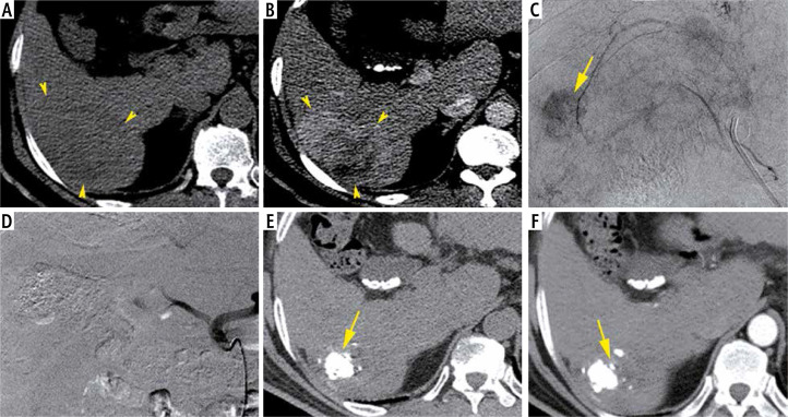
Fig. 1.
A 59-year-old man with chronic hepatitis C and raised AFP (8513 ng/ml), Child-Pugh class A6, MELD score of 12 and BCLC stage C. Axial non-enhanced CT image (A) show a slightly hypodense lesion of the segment VI with heterogeneous enhancement on the arterial phase (B). The HCC size is about 80 mm. Hyperselective TACE demonstrate a nodular blush (C) which completely disappeared after chemoembolization (D). CT control in axial plan performed 6 weeks later shows retraction of the HCC and intense Lipiodol fixation clearly identified on the non-enhanced CT (E) without any enhancement on the arterial phase (F)

