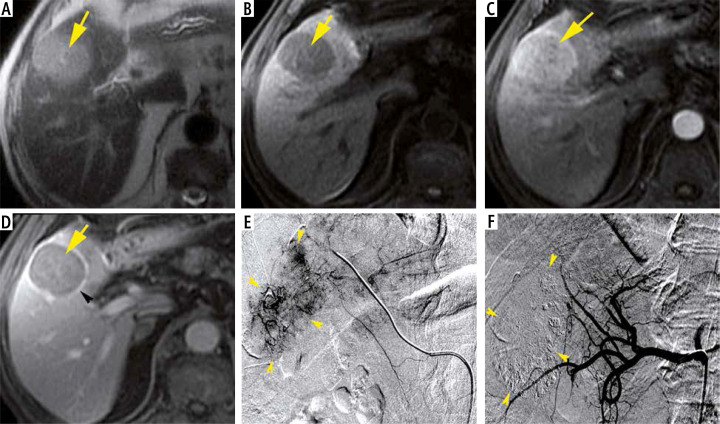
Fig. 3.
An 80-year-old man with chronic hepatitis C and raised AFP (845 ng/ml), Child-Pugh class A6, MELD score of 9 and BCLC stage C. Axial MRI T1 FS images demonstrate a rounded HCC (yellow arrows) of segment V, showing hyposignal on the non-enhanced image (A), with homogeneous wash-in at arterial phase compared to adjacent normal liver (B) and wash-out on portal venous phase (C). Note the enhancing capsule (arrow-head) in portal venous phase (C). The HCC size is about 38 mm. DSA images demonstrate tumoral blush (arrow-heads) (D, E). Note the total extinction of tumoral blush after Lipiodol-doxorubicin injection (arrow-heads) (F)

