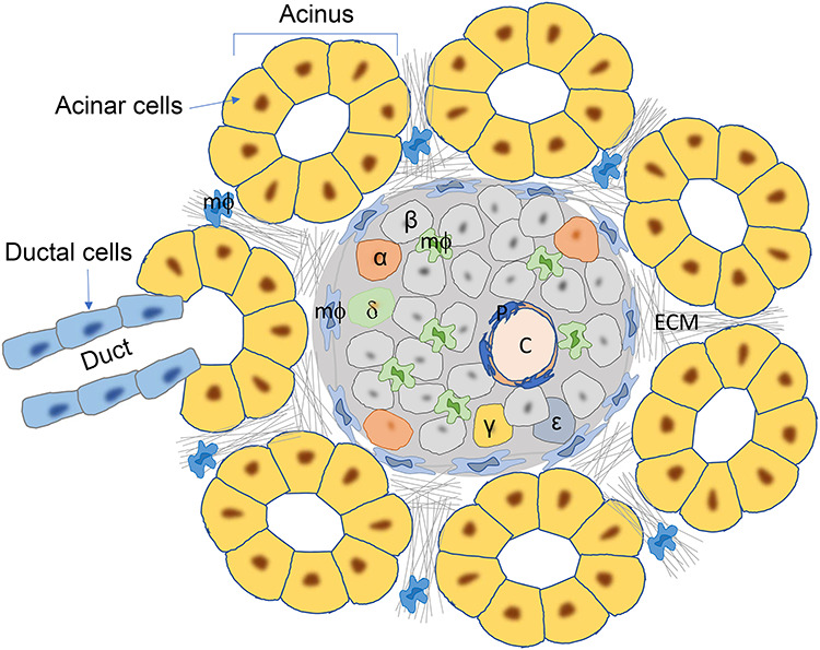
Figure 1.

Pancreas histology at steady state, shown by micrographic view of the mouse pancreas. The islet of Langerhans is surrounded by pancreatic acini, which are composed of exocrine acinar cells. The acinus is also connected to exocrine duct, which is formed by ductal cells. The islet contains various types of endocrine cells, depicted by different colors and labeled (α, β, δ, γ, and  ). The islet capillaries (C) are coated by pericytes (P). Heterogeneous populations of macrophages (mФ) are seen in both exocrine pancreas and the islet. ECM is found in all area of the pancreas, including the exocrine–endocrine interfaces.
