Figure 4.
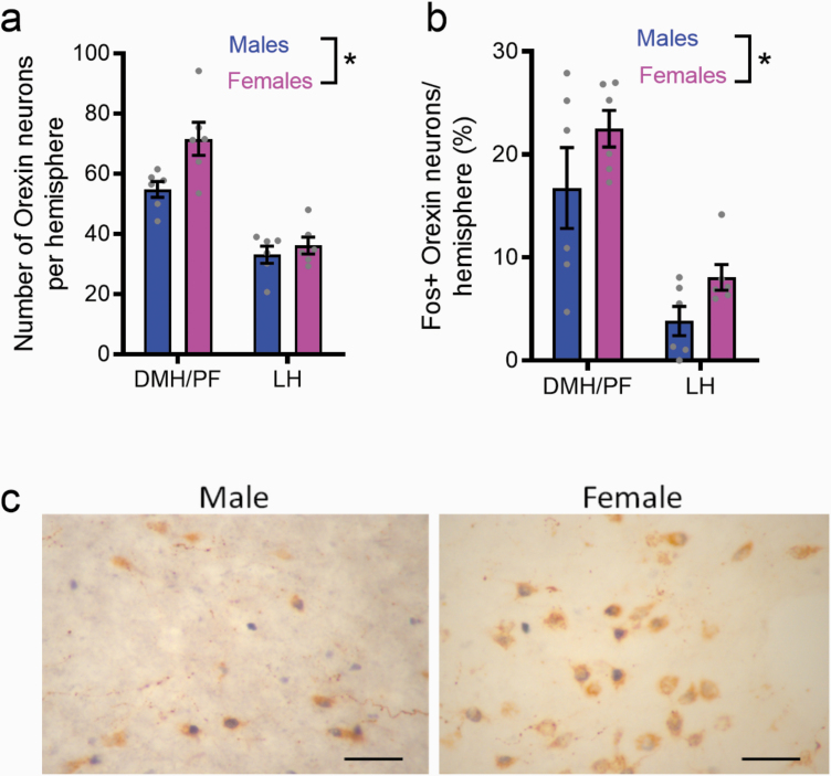
Higher number and activity of orexin-expressing cells in female rats. Immunohistochemical studies revealed that female rats had a higher number of orexin-expressing neurons across the dorsomedial hypothalamus/perifornical (DMH/PF) and lateral hypothalamus (LH) of the hypothalamus compared with males (a). Females also had an increased percentage of orexin cells that expressed cFos in the hypothalamus compared with males (b). Representative micrographs (40×) for males and females are presented (c). Orexin label is brown, cFos is black. Scale bar = 50 μm. *P < .05.

