FIGURE 4.

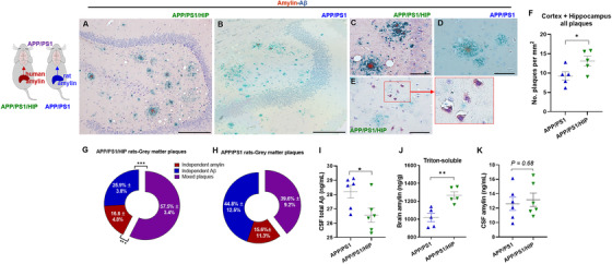
Amylin–amyloid beta (Aβ) cross‐seeding and impaired cerebrospinal fluid (CSF)–brain Aβ exchange by high blood amylin levels (both rat and human amylin) in APP/PS1 rats. A‐E, Representative immunohistological images of co‐staining with anti‐amylin (brown) and anti‐Aβ (green) antibodies in brain sections from 16‐month‐old APP/PS1/HIP rats (A, C, E) and APP/PS1 rats (B, D; n = 5 rats/group). Amylin–Aβ plaques (A‐D) and amylin depositions within neurons (E) are shown. F, Analysis of total plaques including Aβ plaque, amylin plaque, and amylin‐core Aβ plaque depositions in APP/PS1 and APP/PS1/HIP rat brains, assessed from immunohistological images (n = 5 rats/group; data were normalized to total imaging area). G, H, Relative distribution of amylin, Aβ, and amylin–Aβ in plaques in the gray matter of APP/PS1/HIP rat brains (G; n = 5) and APP/PS1 rat brains (H; n = 5). I‐K, CSF total Aβ (I; n = 6 rats/group), brain amylin (Triton‐soluble fractions of tissue homogenate; n = 5 rats/group; J), and CSF amylin (K; n = 6 rats/group) levels in APP/PS1/HIP versus APP/PS1 rats at 16 months of age. Scale bars, 200 μm (A, B), 50 μm (C‐E). Data are means ± standard error of the mean; P ≤ 0.05 *, P ≤ 0.01 **, P ≤ 0.001 ***; two‐tailed, unpaired t test
