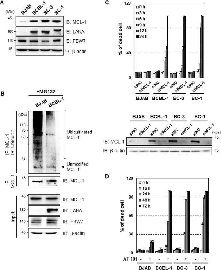
Fig 6. MCL-1 is essential for PEL cell survival.
(A) KSHV- BJAB cell, KSHV+ PEL cell (BCBL-1 and BC-3), and KSHV+EBV+ PEL cell (BC-1) lysates were subjected to IB with anti-LANA and anti-MCL-1 antibodies. β-actin is shown as a loading control. (B) The cells were treated with MG132 (10 μM), followed by IP with an anti-MCL-1 antibody and IB with an anti-ubiquitin antibody. (C) BJAB and KSHV-infected PEL cell lines were transfected with negative control siRNA or siRNA that target MCL-1 (100 pmol) for the indicated periods of time, followed by staining with trypan blue solution. The effect on MCL-1 protein was determined by counting of trypan blue-stained cells. Cells were transfected with scrambled or MCL-1 siRNA for 12 h, followed by immunoblotting (IB) with anti-MCL-1 or anti-β antibodies. (D) Cells were treated with 10 μM AT-101 (MCL-1 inhibitor) for the indicated periods of time and then stained with trypan blue solution.

