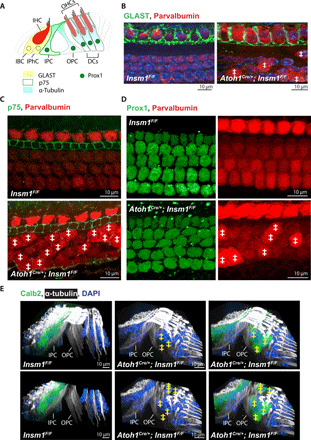
Fig. 2. oc-IHCs are surrounded by outer-compartment, and not inner-compartment, supporting cells.

(A) Supporting cells in the organ of Corti are identified by their expression of distinct markers, including GLAST (IBCs and IPhCs), p75 (IPCs), Prox1 (in the nuclei of IPCs, OPCs, and DCs), and α-tubulin (IPCs, OPCs, and DCs). (B to D) Top view of middle turns from P0 control and Insm1 cKO cochlea, with GLAST labeling IBCs and IPhCs in (B), p75 labeling IPCs in (C), and Prox1 labeling the nuclei of IPCs, OPCs, and the three rows of DCs in (D). Parvalbumin labels IHCs, oc-IHCs, and OHCs (with brighter expression in IHCs and oc-IHCs, where ‡ denotes oc-IHC). (E) Side views of middle turns from control and cKO adult cochlea, with α-tubulin labeling the IPCs, OPCs, and DCs. IPCs and OPCs separate cells in the inner compartment from those in the outer compartment in both control and mutant cochlea. The lower panel has the top sections removed to better visualize the IPC and OPC columnar processes. Calb2 labels IHCs and oc-IHCs (denoted with ‡). DAPI, 4′,6-diamidino-2-phenylindole.
