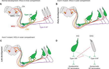
Fig. 8. Schematic illustration and hierarchical model of organ of Corti innervation.

(A) In mature control cochleae, LOC efferents (orange) make axodendritic connections to type I afferent fibers (green) projecting to IHCs (green). Type II afferents (purple) cross into the outer compartment, turn basally, and send collateral projections to a number of OHCs (gray). MOC efferents (red) make axosomatic connections with OHCs. The MSO, LSO, and cochlear nucleus (CN) are indicated. (B) In Insm1 cKO cochleae, type I afferents (AF) cross into the outer compartment to innervate oc-IHCs when there are no interposed OHCs. MOCs make axodendritic connections with type I afferents projecting to these oc-IHCs. oc-IHCs not innervated by type I afferents instead receive collateral projections from type II afferents. These cells also maintain axosomatic connection with MOC fibers, similar to surrounding OHCs. Over the course of development, many type II connections with oc-IHCs are lost, resulting in oc-IHCs not innervated by type I or II afferents (C). These cells also lose efferent connections. Hence, efferent termination depends on the type of afferent fiber connecting the HCs. (D) Hierarchical logic for the development of the organ of Corti innervation: Barb-ended lines (green) denote signaling that attracts, or is permissive to, innervation. T-ended lines (red) denote signaling that repels, or is nonpermissive to, innervation. IHCs attract/permit, and OHCs repel, innervation by type I afferents. Both IHCs and OHCs attract/permit innervation by type II afferents, but type I afferent innervation supersedes that of type IIs. Type I afferents induce axodendritic innervation by efferents (EF), whereas type II afferents innervation is required to establish and maintain axosomatic efferent innervation on the hair cell.
