Fig. 5.
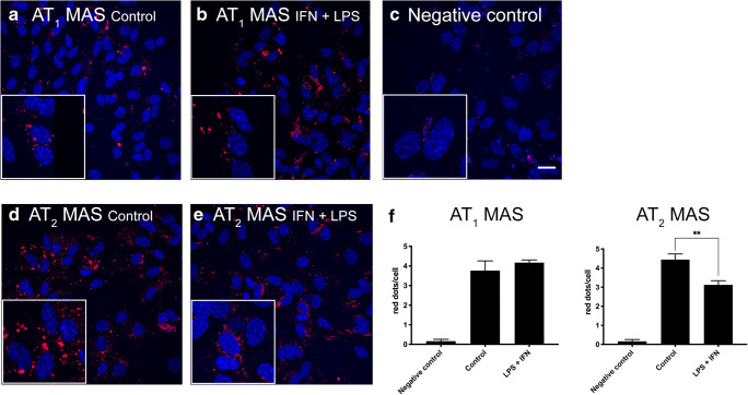
AT1-MasHet and AT2-MasHet expression in microglial primary cultures treated with LPS and IFN-γ. A–E Expression of AT1R-MasR and AT2R-MasR heteromers in primary microglial cultures was determined by proximity ligation assay (PLA), which was performed using specific primary antibodies against AT1, AT2, and Mas receptors. Confocal microscopy images (stacks of 4 consecutive planes) show heteroreceptor complexes; nuclei are Hoechst-stained (blue). Microglial cultures were incubated for 48 h in the absence (A, C, D) or in the presence (B, E) of 1 μM LPS and 200 U/mL IFN-γ. Scale bar: 20 μm. F: Bar graph showing the percentage of AT1R-MasR and AT2R-MasR clusters as red dots/cell. **p < 0.01; Student’s t test versus resting cells

