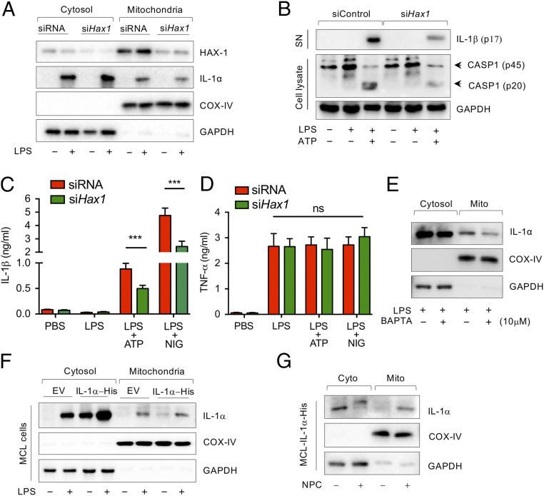
Fig. 3.
LPS-induced pro-IL-1α mitochondrial localization is Hax1- and Ca2+-dependent. (A) WT peritoneal macrophages were transfected with siControl or siHax1 for 48 h and stimulated with PBS or LPS for 3 h. Immunoblotting analysis for HAX-1, IL-1α, COX-IV, and GAPDH in cytosolic and mitochondrial fractions. (B) WT peritoneal macrophages were transfected with siControl or siHax1 for 48 h and stimulated with PBS or LPS for 3 h before ATP (2 mM) treatment for 30 min. Immunoblotting analysis for IL-1b in supernatant (SN), caspase-1, and GAPDH in cell lysate. (C and D) WT peritoneal macrophages were transfected with control siRNA or siHax1 for 48 h and stimulated with PBS or LPS for 3 h before ATP (2 mM) treatment for 30 min. IL-1β (C) and TNF-α (D) production were measured by ELISA. (E) WT macrophages were stimulated with LPS for 1 h and then treated with or without BAPTA-AM for 3 h. Immunoblot analysis for IL-1α, COX-IV, and GAPDH in cytosolic and mitochondrial fraction. (F) IL-1α-His stable expressing MCL cells were treated with or without LPS for 3 h. Immunoblot analysis for IL-1α, COX-IV, and GAPDH in cytosolic and mitochondrial fractions. (G) IL-1α-His stable expressing MCL cells were treated with or without NPC for 3 h. Immunoblot analysis for IL-1α, COX-IV, and GAPDH in cytosolic and mitochondrial fractions. Immunoblots are representative of three independent experiments shown in A, B, and E–G. All data represent mean ± SD. Student’s t test was used for (C and D). ***P < 0.001.

