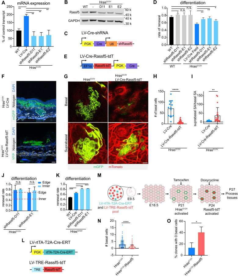
Figure 5. Rassf5 is a necessary and sufficient driver of HrasG12V-induced differentiation.
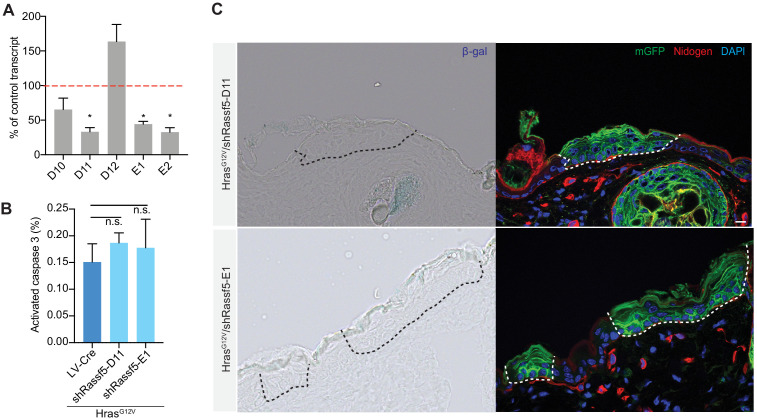
(A) Rassf5 mRNA is increased in isolated HrasG12V E18.5 basal cells. shRNAs targeting Rassf5 efficiently knock down transcript expression. n = 3 animals per genotype. (B) Immunoblot of Rassf5 expression in HrasG12V E18.5 epidermis. (C) LV-Cre-shRassf5 construct for simultaneous knock down of Rassf5 and induction of HrasG12V expression. (D) Quantification of differentiation using EdU-BrdU pulse/chase differentiation assay. Depletion of Rassf5 promotes renewal in HrasG12V epidermis. At least 75 cells were counted per animal. n ≥ 3 animals per genotype. (E) Diagram of construct containing Rassf5 open-reading frame (ORF) fused with tdTomato (Rassf5-tdT) and co-expressing Cre. (F) Representative images of HrasG12V clone and HrasG12V/Rassf5-tdT clone in E18.5 epidermis. Overexpression of Rassf5 yields clones with reduced basal compartment and extensive suprabasal compartment. Scale bar is 25 µm. (G) Whole mount images of HrasG12V clone and HrasG12V/Rassf5-tdT clone in E18.5 epidermis. Scale bar is 25 µm. (H) Quantification of basal cell numbers in E18.5 HrasG12V and HrasG12V/Rassf5-tdT clones. Twenty-two clones were counted in total. n = 3 animals per genotype. (I) Quantification of the ratio of suprabasal surface area to basal surface area of HrasG12V and HrasG12V/Rassf5-tdT clones in E18.5 epidermis. Twenty-two clones were counted in total. n = 3 animals per genotype. (J) Difference between the renewal rate of edge and inner cells is lost in HrasG12V/shRassf5 clones. Animals are >10 weeks old. Red dash lines represent renewal rate of edge and inner cells from week 10 clones. At least 120 cells were counted per genotype. n = 3 animals. (K) Knock down of Rassf5 in adult HrasG12V epidermis significantly increases renewal rate. At least 75 cells were quantified per animal. n ≥ 3 animals per genotype. (L) LV-rtTA-T2A-CreER and LV-TRE-Rassf5-tdT constructs for inducible expression of HrasG12V and Rassf5. (M) Schematic of Rassf5 overexpression in adult HrasG12V clones. Developing embryos were broadly transduced with LV-rtTA-T2A-CreER and sporadically transduced with LV-TRE-Rassf5-tdT. At P21, tamoxifen injection induced expression of HrasG12V, and at P24, doxycycline injection induced expression of Rassf5. Tissues were processed at P27. (N) Clones expressing HrasG12V/Rassf5 have a reduced basal cell population compared to clones expressing HrasG12V. n = 3 animals per condition. A total of 200 clones per condition were counted. (O) There are proportionally fewer HrasG12V clones composed of only differentiated cells compared to HrasG12V/Rassf5. At least 59 clones were counted per animal. n = 3 animals per condition. For (H,I,N), each dot represents an individual clone. For (A,D,H–K,N,O), the center line represents the mean and errors bars the s.d. Two-tailed Student’s t-tests were used. n.s. denotes p value > 0.05. * denotes p value < 0.05; ** denotes p value < 0.01*** denotes p value < 0.001; **** denotes p value < 0.0001.


