Fig. 3.
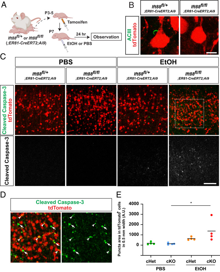
Neuronal cilia suppress Caspase-3 activation in EtOH-exposed cerebral cortex. (A) Experimental scheme. (B) tdTomato-labeled layer V neurons have ciliary axonemes (green) in ER81-lineage neuron-specific Ift88 conditional heterozygous mice but not in conditional homozygous knockout mice. (Scale bar, 10 μm.) (C) Activated Caspase-3 (green or white) within tdTomato-labeled (red) cortical layer V in M1 in the indicated genotype × treatment groups 24 h postexposure, showing augmented Caspase-3 activation in EtOH-exposed conditional homozygous knockout mice. (Scale bar, 100 μm.) (D) Higher magnification view of boxed area in C, showing augmented caspase activation in tdTomato-positive neurons (arrowheads) as well as in tdTomato-negative surrounding cells (arrows). (E) Quantification of activated Caspase-3 puncta within TdTomato-positive neurons in layer V in M1. *P < 0.05 by post hoc Tukey test following two-way ANOVA.

