Fig. 4.
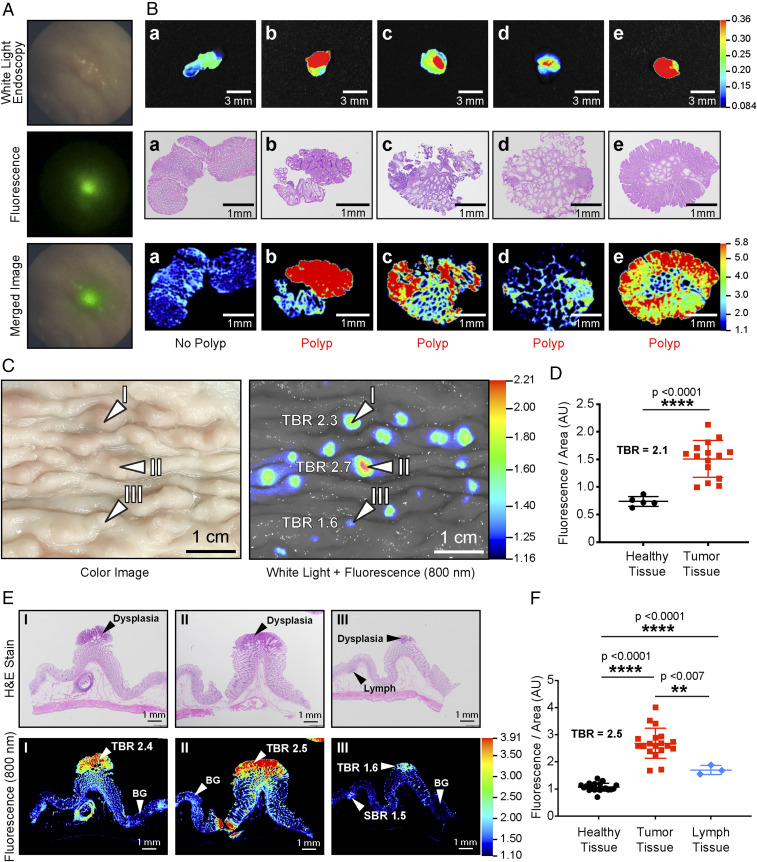
NIRF-guided biopsy in porcine model of colorectal carcinogenesis. (A) NIRF-highlighted polyp in the colon of an APC1311/+ pig (80 kg) after injection with 6QC-ICG (1.0 mg/kg). (B) (Top row) Wide-field NIRF images of tissues obtained during NIRF-guided biopsy. (Middle row) Corresponding H&E-stained tissue of biopsied tissue confirms the histological status of the tissues. (C) Images and tissue scans of colon tissue from an APC1311/+ porcine model injected with 6QC-ICG (euthanized at 24 h PI, 1.0 mg/kg). Wide-field imaging with white light (Left) and corresponding 800-nm fluorescence image (Right). (D) Quantification of fluorescence of healthy and tumor tissues from wide-field imager for APC1311/+ pigs injected with 1.0 mg/kg of probe. (E) Colon tissue slices stained with H&E (Top) and their corresponding flat-bed NIRF imager 800-nm scans (Bottom). (F) Quantification of fluorescence signals of healthy, tumor, and lymph sections of scanned colon tissues taken from the APC1311/+ pigs injected with 1.0 mg/kg of probe. Error bars represent SDs.

