Fig. 5.
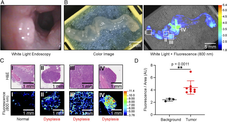
Imaging colorectal cancer injected with 6QC-ICG in AOM-DSS mice. (A) Screen capture of colon of an AOM-DSS–treated mouse in white-light endoscopy. (B) Images of colon tissue from an AOM-DSS mouse model injected with 6QC-ICG (euthanized 14 h PI, 2.3 mg/kg). Color image (Left) and white light + 800 nm fluorescence (Right). (C) H&E-stained slide images of corresponding tissue slices (Top) and flat-bed fluorescence scanner 800-nm scans of H&E-stained slides (Bottom). (D) Quantification of fluorescence signals of background and tumor sections of scanned colon tissue taken from AOM-DSS–treated mice (n = 5) (euthanized14 h PI, 2.3 mg/kg). Error bars represent SD.

