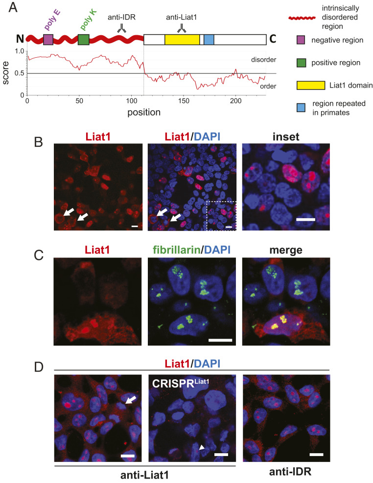
Fig. 1.
Conserved features of Liat1 and its subcellular localization. (A) Schematic of the mouse Liat1 protein showing evolutionarily conserved features (indicated in Right). Shown are the relative locations of anti-IDR and anti-Liat1 antibody recognition. Also shown is an IUPred2A plot revealing that the N-terminal half of Liat1 comprises an IDR (41). (B) Anti-Liat1 immunocytochemistry of HEK293T cells transfected with plasmids expressing mouse 3xHALiat1. (C) Colocalization of Liat1 with fibrillarin, a marker of the DFC of the nucleolus. (D) Anti-Liat1 immunocytochemistry of nontransfected HEK293T cells (Left). Also probed was a mixed population of cells transfected with CRISPRLIAT1-B, targeting the portion of LIAT1 encoding the repeated motif. The arrowhead indicates Liat1-containing nucleoli that could be seen in some cells (Center). Anti-IDR immunocytochemistry of nontransfected HEK293T cells (Right). White arrows, mitotic cells. (Scale bars: 10 µm.)

