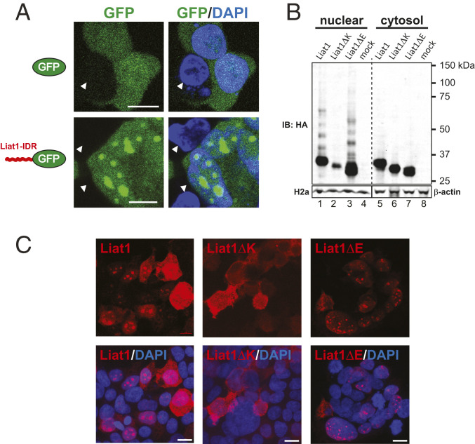
Fig. 4.
The IDR of Liat1 facilitates LLPS and contains a poly-K nucleolar targeting signal. (A) Direct fluorescence of eGFP (GFP; Upper) and of eGFP bearing an N-terminal fusion of the Liat1 IDR (Lower). Arrowheads indicate nontranfected cells. (Scale bars: 10 µm.) (B) Anti-HA immunoblot of the nuclear and cytosolic fractions of HEK293T cells expressing wild-type 3xHALiat1 or Liat1 lacking the poly-E region (Liat1ΔE) or the poly-K region (Liat1ΔK; Upper). Anti-Histone H2a and anti–β-actin of the nuclear and cytosolic fractions, respectively (Lower). IB: immunoblot using the indicated antibody. (C) Anti-Liat1 immunocytochemistry of cells expressing wild-type Liat1, Liat1ΔK, or Liat1ΔE. (Scale bars: 15 µm.)

